


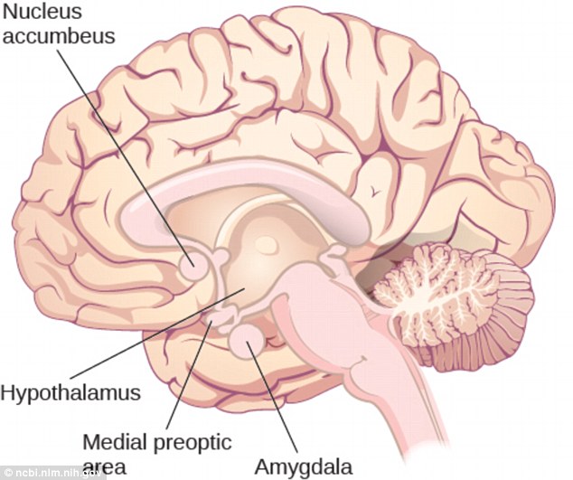
The study traced the trigger of attraction to the hypothalamus, an evolutionarily ancient structure at the bottom-center of the brain that`s responsible for controlling major bodily functions such as body temperature, hunger, thirst and sexual behavior.
The researchers at the University of North Carolina (UNC) School of Medicine examined the medial preoptic area (mPOA) of the brain - a specific set of neurons inside the hypothalamus.
Previous research found the mPOA was important for social and reproductive behaviour in all vertebrate species studied from fish to human.
But until now it has been unclear whether this area drives social motivation through circuit connections with reward systems in the brain.
To test how this brain area works in attraction, the researchers exposed female mice to male mouse urine.
When female mice were exposed to the odor of male mouse urine, a large number of the neurons in the mPOA became excited into greater activity.
The researchers also found that these neurons responded more strongly to male mouse urine when females had high circulating levels of estrogen or a combination of estrogen/progesterone, which surges before the mice become fertile.
By contrast, the same neurons were not excited when the mice were exposed to the odor of female mouse urine or attractive odors like food.
Dr Jenna McHenry, a postdoctoral research associate at UNC and first author of the research paper published in Nature Neuroscience, said: `This suggests that certain neurons in the brain may be specialized to prefer social rewards over nonsocial rewards, and that the processing of social cues is sensitive to circulating hormones.`
Normal microscopy techniques only allow imaging of brain cells in awake mice for brain cells that are a fraction of a millimeter below the brain`s surface.
But the mPOA is several millimeters deep in the brain, so to visualize those neurons the researchers had to use a special microscopy technique.
Dr Garret D Stuber, associate professor of psychiatry, cell biology & physiology at UNC and leader of the study said him and his team used tiny tubular lenses connected from their microscope to the mPOA of the mouse`s brain.
With a technique known as two-photon calcium imaging, the scientists were able to visualize the activity of mPOA neurons in awake female mice.
To enhance the accuracy of the technique, the researchers used mice that had been genetically engineered so that only their mPOA neurons could be imaged.
The researchers examined the medial preoptic area (mPOA) of the brain - a specific set of neurons inside the hypothalamus. Previous research found the mPOA was important for social and reproductive behavior in all vertebrate species studied from fish to human
Dr Stuber said: `With our setup, we could image the mice a couple of times a week and each time find the same cells that we previously recorded brain activity from.`
The researchers also used optogenetics to demonstrate the effect of stimulating the mPOA area.
Optogenetics (from Greek optos, meaning `visible`) uses light to control neurons which have been genetically sensitized to light.
It involves introducing light-sensitive proteins into cells, which when activated, can affect the cell`s activity.
They found that stimulating the mPOA neurons with light triggered the release of the neurotransmitter dopamine from a part of the brain called the ventral tegmental area (VTA).
Dopamine helps control the brain`s reward and pleasure centers.
Both male and female mice whose mPOA neurons were stimulated moved closer to mice of the opposite sex.
Dr Stuber said: `On the whole, the data suggest that these mPOA neurons help drive social attraction toward a potential mate.`
Stuber`s team plans to follow up by applying similar imaging and optogenetics methods to the workings of the VTA.
The findings also have implications for anxiety, depression and related disorders, which can be triggered or worsened in some women by shifts in hormone levels.
Dr McHenry said: `While hormone-related changes in motivation are important for mating or maternal behaviour in female mice, some atypical hormonal changes in women appear to underlie reproductive mood disorders, such as postpartum depression.`
Studying these hormone-sensitive circuits that control motivational states could lead to new drugs to target these mood disorders.
USING OPTOGENETICS TO ALTER BRAIN CELLS
Optogenetics (from Greek optos, meaning `visible`) uses light to control neurons which have been genetically sensitized to light.
The technique, known as optogenetics, was pioneered by Karl Diesseroth at Stanford University, is a new way to manipulate and study nerve cells using light.
The techniques are rapidly becoming the standard method for investigating brain function.
It involves introducing light-sensitive proteins into cells, which when activated, can affect the cell`s activity.
Experiments in rats have shown that when neurons are altered with these protein complexes and targeted with light – typically using fibre optics to deliver laser pulses – the proteins become activated and change the activity of the cell, effectively switching it on or off depending on the proteins introduced.
This provides an invaluable method for scientists to peer into the complex network of cells to see which regions are implicated in different scenarios.
But the approach also has great potential for treatment, where the activity of specific sets of brain cells could be tweaked using light.
/Daily Mail/


-1780923278.jpg&h=190&w=280&zc=1&q=100)