It's been done in Cardiff, Nottingham, Cambridge and Stockport, as well as London England and London Ontario.
Doctors hope it will help increase understanding of a range of neurological disorders and could be used instead of invasive biopsies.
I volunteered for the project - not the first time my brain has been scanned.
Computer games
In 2006, it was a particular honour to be scanned by the late Sir Peter Mansfield, who shared a Nobel prize for his work on developing Magnetic Resonance Imaging, one of the most important breakthroughs in medicine.
He scanned me using Nottingham University's powerful new 7 Tesla scanner. When we looked at the crisp, high resolution images, he told me: "I'm a physicist, so don't ask me to tell you to whether there's anything amiss with your brain - you'd need a neurologist for that."
I was the first UK Biobank volunteer to have their brain and other organs imaged as part of the world's biggest scanning project.
More recently, I had my brain scanned while playing computer games, as part of research into the effects of sleep deprivation on cognition.
So my visit to the Cardiff University's Brain Research Imaging Centre (CUBRIC) held no particular concerns.
The scan took around 45 minutes and seemed unremarkable.
A neurologist was on hand to reassure me my brain looked normal.
My family quipped that they were happy that a brain had been found inside my thick skull.
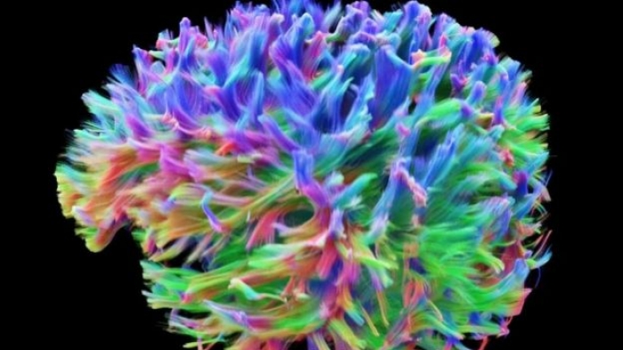
But nothing could have prepared me for the spectacular images produced by the team at Cardiff, along with engineers from Siemens in Germany and the United States.
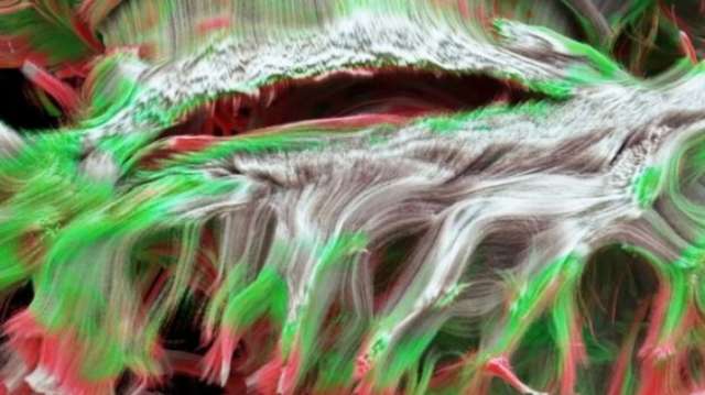
The scan shows fibres in my white matter called axons. These are the brain's wiring, which carry billions of electrical signals.
Axonal density
Not only does the scan show the direction of the messaging, but also the density of the brain's wiring.
Another volunteer to be scanned was Sian Rowlands who has multiple sclerosis.
Like me, she is used to seeing images of her brain, but found the new scan "amazing".
Conventional scans clearly show lesions - areas of damage - in the brain of MS patients.
But this advanced scan, showing axonal density, can help explain how the lesions affect motor and cognitive pathways - which can trigger Sian's movement problems and extreme fatigue.

The scan shows fibres in the brain's white matter, called axons
Prof Derek Jones, CUBRIC's director, said it was like getting hold of the Hubble telescope when you've been using binoculars.
"The promise for researchers is that we can start to look at structure and function together for the first time," he said.
The extraordinary images produced in Cardiff are the result of a special MRI scanner - one of only three in the world.
The scanner itself is not especially powerful, but its ability to vary its magnetic field rapidly with position means the scientists can map the wires - the axons - so thinly it would take 50 of them to match the thickness of a human hair.
The scanner is being used for research into many neurological conditions including MS, schizophrenia, dementia and epilepsy.
My thanks to Sian, Derek and all the team at CUBRIC.
More about: #brain


