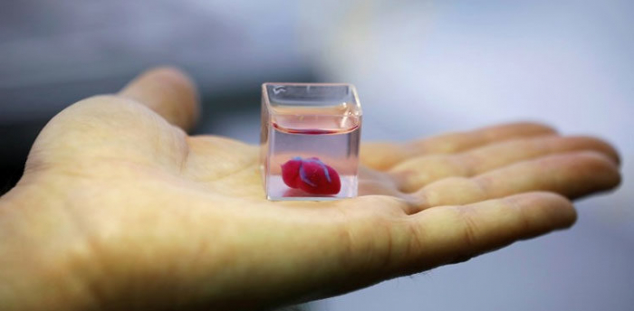
Their method, which was described in the journal Science, replicates the body's own complex biological scaffolds using its most abundant protein at the highest level of precision yet achieved.
The structures are then further embedded with living cells and capillaries at a resolution of 20 micrometers, far greater than most 3D printers used to create plastic structures.
"What we were able to show was you can actually 3D print a heart valve out of collagen," Adam Feinberg, a co-author of the paper who is a professor of biomedical engineering at Carnegie Mellon University, told AFP.
"We have not yet put them in an animal but we built a benchtop system that can simulate the pressure and the flow rate of the human body, and we show that we put it in there and it works."
The team used MRI scans of human hearts to reproduce patient-specific parts, which achieved outcomes like synchronized beating and opening and closing of valves.
In April, an Israeli team unveiled a 3D print of a heart with human tissue and vessels, but the organ did not have the ability to pump.
- Open-source design -
Previous attempts at printing the scaffolds, known as extracellular matrices, had been hindered by limitations that resulted in poor tissue fidelity and low resolutions.
Collagen, which is an ideal biomaterial for the task since it is found in every tissue of the human body, starts out as a fluid and attempting to print it resulted in puddle of Jell-O-like material.
But the scientists were able to overcome these hurdles by using rapid changes in pH to cause the collagen to solidify with precise control.
"That's the very first version of a valve, and so anything that we engineer as a product will actually get better and better," Feinberg said.
The technique, known as Freeform Reversible Embedding of Suspended Hydrogels (FRESH), allows the collagen to be added layer by layer in a support bath of gel, which is then melted away by heating it from room to body temperature, leaving the structure undamaged.
The team's designs are open-source, meaning other labs can replicate the results and print the same parts.
- Patching organs -
In a commentary that appeared in Science, biomedical engineers Queeny Dasgupta and Lauren Black, who are at Tufts University and were not involved with the research, wrote: "Other methods of printing vasculature or printing collagen have been demonstrated but did not achieve the precision or resolution" of the new method.
They added that the new technique creates structures "that substantially increase cell viability" and angiogenesis, the formation of new blood vessels.
But they cautioned that improvements in functional outcomes are still needed and the ultimate goal would be an even greater resolution of 1 micrometer.
In the long term, the technique could one day be used to print viable hearts or other organs. There are 4,000 people in the United States awaiting a heart transplant and millions of others worldwide who need hearts but may be ineligible.
Challenges to that goal include the generation of the billions of cells needed to bioprint large tissues, achieving manufacturing scale, and following regulatory processes so that it can be tested in animals and eventually humans.
"I think more near term is probably patching an existing organ," such as a heart that has suffered a loss of function through a heart attack, or a damaged liver, said Feinberg.
More about: science heart human-tissue


